
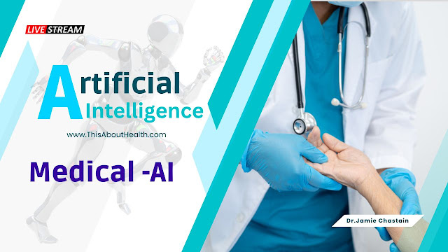
Role of Artificial Intelligence in the Medical Field
 |
| Artificial Intelligence Is Changing Medical Imaging to Improve Patient Care |
Imaging is additionally evolving from its initial focus—diagnosing medical conditions—to half in} associate degree integral part in treatment still, particularly within the space of cancer. Doctors area unit setting out to rest on imaging to assist them to monitor tumors and therefore the unfold of cancer cells so they need a much better, quicker means of knowing if therapies area unit operating. That new role for imaging can remodel the categories of treatments patients can receive, and immensely improve the knowledge doctors get around however well they’re operating, so they will ultimately create higher selections regarding what treatment choices they have.Imaging in medication currently involves subtle ways of analyzing each piece of information to differentiate sickness from health and signal from noise. If the primary few decades of radiology were regarding purification the resolution of the images taken of the body, then the following decades are dedicated to deciphering that information to make sure nothing is unnoticed.
In consecutive 5 years, we are going to see practical imaging become a part of care,” says Dr. Basak Dogan, prof of radiology at the University of Texas Southwestern heart. “We don’t see the present commonplace imaging respondent the $64000 clinical queries. however practical techniques are the solution for patients WHO wish higher exactitude in their care so that they will build higher awareness selections.
Detecting problems earlier
The first hurdle in creating the foremost of what pictures will offer whether they're X-rays, X-radiation (CT) scans, resonance imaging (MRI), or ultrasounds is to alter the reading of them the maximum amount as doable, that saves radiologists valuable time. Computer-aided algorithms have established their value during this space, as huge computing power has created it doable to coach computers to tell apart abnormal from traditional findings. code specialists and radiologists are teaming up for years to come back up with these formulas; radiologists feed pc programs their findings on tens of thousands of traditional and abnormal pictures, which teaches the pc to tell apart once pictures contain things that fall outside of traditional parameters. The additional pictures the pc needs to compare and learn from, the higher it becomes at fine-tuning the distinctions.
For the U.S. Food associated Drug Administration (FDA) to approve an algorithmic program involving imaging, it should be correct eightieth to ninetieth of the time. So far, the government agency has approved 420 of those for varied diseases (mostly cancer). The government agency still needs that an individual is the final word arbiter of what the machine-learning algorithmic program finds, however such techniques area unit crucial for drooping pictures that may contain suspicious findings for doctors to review—and ultimately give quick answers for patients.
At Mass General Brigham, doctors use fifty such algorithms to assist them with patient care, starting from sleuthing aneurysms and cancers to recognizing embolisms and signs of stroke among emergency-room patients, several of whom can gift with general symptoms that these conditions share. regarding 0.5 are approved by the government agency, and therefore the remaining ones area unit being tested in patient care.
“The goal is to search out things early. In some cases, it's going to take humans days to search out AN correct designation, whereas computers will run while not sleep endlessly and notice those patients World Health Organization want care promptly,” says Dr. Keith Dreyer, chief information science officer and chairperson of radiology at Mass General Brigham. “If we will use computers to try to do that, then it gets that patient to treatment abundant quicker.
Tracking patients more thoroughly
While computer-assisted stringing is the opening move in the desegregation of AI-based support in medication machine learning is additionally turning into a strong thanks to monitoring patients and tracking even the littlest changes in their conditions. this can be particularly essential in cancer wherever the tedious task of deciding whether or not someone’s growth is growing, shrinking, or remaining identical is important for creating choices regarding however well treatments area unit operating. We have hassle understanding what's happening to the growth as patients bear therapy says Dogan. Our customary imaging techniques sadly can’t observe any modification till once midway through chemo which is often months into the process when some quite a shrinkage starts occurring.
Imaging is often helpful in those things by learning changes in tumors that aren’t associated with their size or anatomy. “In the early stages of therapy, most of the changes during a tumor don't seem to be quite at the amount of necrosis,” says Dogan. “The changes area unit associated with modifying interactions between the body’s immune cells and cancer cells.” And in several cases, cancer doesn’t shrink in a certain manner from the skin in. Instead, pockets of cancer cells inside a tumor could go away, whereas others still thrive, going away the general mass additional pockmarked, sort of a moth-eaten sweater. In fact, as a result of a number of that necrosis connected to inflammation, the dimensions of the tumor could even increase in some cases, even supposing that doesn’t essentially indicate additional neoplastic cell growth. customary imaging presently can’t distinguish abundantly |what proportion|what quantity} of a tumor continues to be alive and the way much is dead. The most remarkably used breast-cancer imaging techniques, diagnostic techniques, and ultrasound, are area units designed instead to choose anatomical options.
At UT Southwestern, Dogan is testing 2 ways in which imaging is accustomed to track useful changes in carcinoma patients. In one, victimization funding from the National Institutes of Health, she is imaging carcinoma patients when one cycle of therapy to choose up slight changes in pressure around the tumor by injecting micro bubbles of gas. Ultrasound measures changes in the pressure of those bubbles, that tend to accumulate around tumors; growing cancers have additional blood vessels to support their growth, compared to different tissues.
In another study, Dogan is testing optoacoustic imaging that turns lightweight into sound signals. Lasers are shone on breast tissue, inflicting cells to oscillate which creates sound waves that are captured and analyzed. this system is similar in temperament to noticing a tumor's gas levels since cancer cells tend to wish for additional gas than traditional cells to continue growing. Changes in sound waves will notice that elements of the growth are still growing and that doesn't seem to be. just by imaging the growth we can will we can tell that is presumably to spread to the humor nodes and that doesn't seem to be says Dogan. Currently, clinicians can’t tell that cancers can unfold to the humor and that won’t. It may provide United States data concerning however the growth goes to behave and probably save patients superfluous node surgeries that are currently a part of customary care.
The technique might additionally facilitate the notice of early signs of cancer cells that have to unfold to alternative components of the body well before they show up in visual scans and while not the requirement for invasive biopsies. that specialize in organs in that cancer cells generally unfold, like the bones, liver, and lungs, might provide doctors an advantage in catching these new deposits of cancer cells.
Spotting unseen abnormalities
With enough information and pictures, these algorithms might even realize aberrations for any condition that no human might observe says Dreyer. His team is additionally engaged in developing an associate algorithmic program that measures bound biomarkers within the frame whether or not anatomical or practical, therefore it will flag changes in those metrics that would recommend somebody is probably going to own a stroke fracture heart failure, or another adverse event. That’s the Holy Grail of imaging says Dreyer, and whereas it’s a couple of years away those square measure the styles of things that square measure about to be transformational in health care for AI.
To get there it'll take tons and a lot of information from many thousands of patients. however, the siloed healthcare systems of the U.S. mean that pooling such data is difficult. united learning within which scientists develop algorithms that square measure applied to completely different institutions’ anonymized patient-information databases is one resolution. That way privacy is maintained and establishments won’t be got to jeopardize their secure systems.
If a lot of these models square measure valid through federate learning or otherwise AI-based imaging might even begin to assist patient reception. As COVID-19 created self-testing and telehealth a lot of routine individuals might eventually be ready to get imaging info through transportable ultrasounds provided via a smartphone app for instance.
The real modification in health care that's about to happen from AI is that it'll deliver loads of solutions to patients themselves or before they become patients so that they'll keep healthy says Dreyer. that might maybe be the foremost potent thanks to optimizing imaging by empowering patients to be told from and create the foremost hep choices potential regarding protective their health.